[無料ダウンロード! √] paranasal sinuses anatomy radiology 340440-Paranasal sinuses anatomy radiographics
· Sphenoid sinuses CT brain (bone windows) Hover on/off image to show/hide findings Tap on/off image to show/hide findings Click image to align with top of page Sphenoid sinuses CT brain (bone windows) The sphenoid sinus can be single or septated into multiple separate sinuses;Paranasal Sinuses MRI examination; · Computed tomography (CT) is currently the modality of choice in the evaluation of the paranasal sinuses and adjacent structures 2 – 7 Its ability to optimally display bone, soft tissue, and air facilitates accurate depiction of anatomy and extent of disease in and around the paranasal sinuses 2 – 5 7 In contrast to standard radiography, CT can clearly show the fine bony anatomy

Paranasal Sinus Anatomy What The Surgeon Needs To Know Intechopen
Paranasal sinuses anatomy radiographics
Paranasal sinuses anatomy radiographics-Anatomy and Physics Sydney Weekend Courses Our Anatomy and Physics weekend courses are designed by surgeons and radiologists to teach you how to effectively study, answer and pass the Surgery GSSE, Radiology Anatomy and Physics Radiology Exams Register for the 21 Anatomy Weekend Course (Sydney, August 21) Register for the 21 Physics · A precise knowledge of the anatomy of the paranasal sinuses is essential for the clinician Conventional radiology does not permit a detailed study of the nasal cavity and paranasal sinuses, and has now largely been replaced by computerised tomographic (CT) imaging This gives an applied anatomical view of the region and the anatomical variants
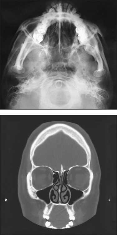


Paranasal Sinuses Radiology Key
/08/19 · Computed tomography (CT) and magnetic resonance imaging (MRI) are the primary imaging modalities for assessment of the paranasal sinuses Both these imaging techniques are far superior to plain radiographs in the evaluation of the sinonasal cavities Plain radiographs of the paranasal sinuses are limited in their ability to delineate the complex bony anatomy or to characterize sinonasalSphenoid sinus clinical significanceAbstract The development of the paranasal sinuses has been well described 1 The maxillary sinuses are the first of the paranasal sinuses to develop;
99 · Radiologic Anatomy of the Paranasal Air Sinuses Sundeep Nayak Over the last decade, significant strides have been made in the imagingguided management of sinonasal inflammation We are now able to achieve an enhanced level of understanding of the complex functional and surgical anatomy of this region, This facilitates meaningful communication withThe paranasal sinuses (latin sinus paranasales) are four bilateral airfilled spaces within bones of the skull surrounding the nasal cavityFour bones of the skull each accommodates a pair of paranasal sinuses that are named according to the bone in which they are locatedThe four sinuses are maxillary sinus,;"Normal CT Anatomy of the Paranasal Sinuses and Nasal cavity" lecture Case discussion (901 min webinar) WEEK 2 // CT Anatomy and normal variants of PNS Reporting on selected cases (67 hour learning time) 9am CET // Live webinar "Anatomy of the Ostiomeatal Complex" lecture Case discussion (901 min webinar) WEEK3
SIMPLIFIED PARANASAL ANATOMY #anatomy #pns #ctpns Also known as antrum of Highmore Largest paranasal sinusPyramidal in shape Base toward the lateral wall ofDevelopment begins in the first trimester of gestation and usually is completed byParanasal sinuses and facial bones radiography Amanda Er (she/her) and Andrew Murphy et al Paranasal sinuses and facial bones radiography is the radiological investigation of the facial bones and paranasal sinuses Plain radiography of the facial bones is still often used in the setting of trauma, postoperative assessments and dental radiography


Maxillary Sinus Radiology Reference Article Radiopaedia Org


Imaging The Paranasal Sinuses Where We Are And Where We Are Going Fatterpekar 08 The Anatomical Record Wiley Online Library
Nov 18, 18 Crosssectionnal anatomy of the head on a cranial CT Scan brain, bones of skull, paranasal sinuses Nov 18, 18 Crosssectionnal anatomy of the head on a cranial CT Scan brain, bones of skull, paranasal sinuses Today Explore When autocomplete results are available use up and down arrows to review and enter to select Touch device users, explore by1 Am J Med Sci 1998 Jul;316(1)212 Functional anatomy and computed tomography imaging of the paranasal sinuses Zinreich SJ(1) Author information (1)Department of Radiology/Otolaryngology, Head & Neck Surgery, The Johns Hopkins Medical Institutions, Baltimore, Maryland 215, USA Standard radiographs are suboptimal in the display of the · Overview of Paranasal Sinus Anatomy The paranasal sinuses consist of paired frontal, ethmoid, maxillary, and sphenoid sinuses The frontal sinuses are located superior to the orbits and ethmoid sinuses Both the superior and posterior walls of the frontal sinus separate the sinus from the cranial vault The floor of the frontal sinus consists of the orbital roof The ethmoid sinuses


Para Nasal Sinuses Anatomy Radiology Students Of A M S Facebook


Www Ijcmr Com Uploads 7 7 4 6 Ijcmr 1967 V4 Pdf
A CT scan of the face produces images that also show a patient's paranasal sinus cavities The paranasal sinuses are hollow, airfilled spaces located within the bones of the face and surrounding the nasal cavity, a system of air channels connecting the nose with the back of the throat There are four pairs of sinuses, each connected to the nasal cavity by small openings View this four minuteMusculoskeletal Ankle Fracture mechanism and Radiography · Surgical Anatomy ofthe Paranasal Sinus M PAIS CLEMENTE The paranasal sinus region is one of the most complex areas of the human body and is consequently very difficult to study The surgical anatomy of the nose and paranasal sinuses is published with great detail in most standard textbooks, but it is the purpose of this chapter to describe


The Preoperative Sinus Ct Avoiding A Close Call With Surgical Complications Radiology
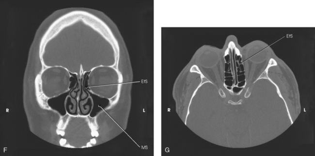


Paranasal Sinuses Radiology Key
· Nasal cavity and paranasal sinuses radiologic anatomy 2 Roadmap Gross Introduction Positioning Anatomy Radiographic X Ray CT Anatomy MRI Questions 4 Introduction Nasal cavity is a passage from the external nose anteriorly to the nasopharynx posteriorly The frontal, ethmoid, sphenoid and maxillary sinuses form the paired paranasalNeck masses Neck Masses in Children; · Tan HM, Chong V (01) CT of the paranasal sinuses normal anatomy, variation and pathology CME Radiology 21–125 Google Scholar Tao Y, Gao Q, Cui Y (1998) The evaluation of paranasal sinus coronal CT scanning in endoscopic sinus surgery Lin Chuang Er Bi Yan Hou Ke Za Zhi 12(8)346 PubMed Google Scholar Underwood AS (1910) An inquiry into the anatomy and


Paranasal Sinus Anatomy What The Surgeon Needs To Know Intechopen


Paranasal Sinuses Radiology And Anatomy Neurosurgery Lounge
· Chief—Head and Neck Radiology, Mount Sinai Medical Center, New York, NY Search for more papers by crosssectional imaging has dramatically changed our approach and understanding of the anatomy and pathology of paranasal sinuses We have moved away from plain film radiographs to modern highresolution sinus computerized tomography (CT) and · title = "Imaging Evaluation of the Paranasal Sinuses", abstract = "Cross sectional imaging has revolutionized the way we approach anatomy and pathology of the paranasal sinus Plain films, once considered standard of care, have been widely replaced by highresolution computed tomography (CT) and magnetic resonance imaging (MRI) · Structures seen Sphenoid,posterior ethmoid and maxillary sinuses(seen best in that order) Zygoma and zygomatic arch Mandible 11 Right and left oblique view 12 Xrays for nasal fractures Lateral view of nasal bones Occlusal view of nasal bone Water's view 13
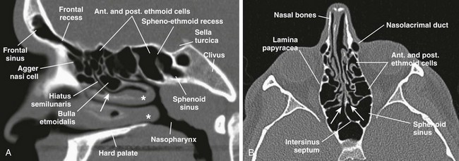


Nose And Sinonasal Cavities Radiology Key
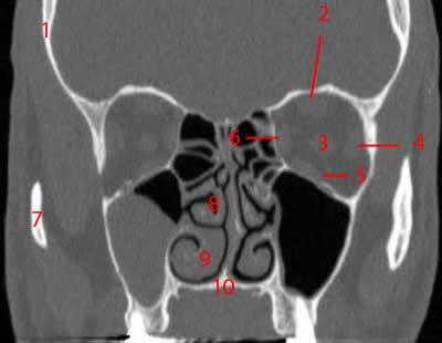


Paranasal Sinuses Ct Anatomy W Radiology
Request PDF Radiology of Paranasal Sinuses Imaging of paranasal sinuses forms an essential part of management of patients with disease of nose and sinuses, and over the last decade withComputed tomography (CT) of the sinuses uses special xray equipment to evaluate the paranasal sinus cavities – hollow, airfilled spaces within the bones of the face surrounding the nasal cavity CT scanning is painless, noninvasive and accurate It's also the most reliable imaging technique for determining if the sinuses are obstructed and the best imaging modality for sinusitisParanasal Sinuses MRI Laurie Loevner and Jennifer Bradshaw Radiology department of the University of Pennsylvania, USA and the radiology department the Medical Centre Alkmaar, the Netherlands
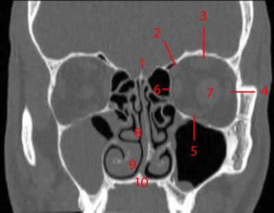


Paranasal Sinuses Ct Anatomy W Radiology
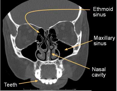


Computed Tomography Ct Sinuses
Computed tomography (CT) scans may help examine paranasal sinuses' anatomy and detect abnormalities (10) Moreover, a CT scan gives a greater definition of the sinuses (11) It is more sensitive than typical radiography tools in detecting sinus pathology, especially within the sphenoid and ethmoid sinuses · The solid knowledge of the surgical anatomy and normal development of the paranasal sinuses is the key element behind achieving superb end results, whether the target is to accomplish a therapeutic surgical procedure or to conduct a clinical trial or research Understanding the surgical landmarks and the anatomical variants of the paranasal sinuses will guide surgeonsEthmoid sinus (known as ethmoidal


Orbits Anatomy Mri Orbits And Paranasal Sinuses Anatomy Free Cross Sectional Anatomy


The Radiology Assistant Paranasal Sinuses
Paranasal Sinuses and Nose Normal Anatomy and Pathologic Processes Authors;/08/15 · It is imperative for all imaging specialists to be familiar with detailed multiplanar CT anatomy of the paranasal sinuses and adjacent structures This article reviews the radiologically relevant embryology of this complex region and discusses the regionspecific CT anatomy of the paranasal sinuses and surrounding structures Radiologists also need to know the clinicalNose and Paranasal Sinuses PD Dr med Basile N Landis Unité de RhinologieOlfactologie Service d'OtoRhinoLaryngologie et de Chirurgie cervicofaciale, Hôpitaux Universitaires de Genève, Suisse Anatomy External Nose Large Nose Thin Nose Huizing, de Groot, Functional reconstructive nasal surgery, 03, Georg Thieme Verlag Huizing, de Groot, Functional reconstructive nasal


Paranasal Sinuses Anatomy In Diagnostic Imaging Wiley Online Library


Paranasal Sinuses Radiology Key
TIRADS TIRADS Thyroid Imaging Reporting and Data System;IPad version of the Radiology Assistant; · With the advent of multidetector computed tomography (MDCT), imaging of paranasal sinuses prior to functional endoscopic sinus surgery (FESS) has become mandatory Multiplanar imaging, particularly coronal reformations, offers precise information regarding the anatomy of the sinuses and its variatio
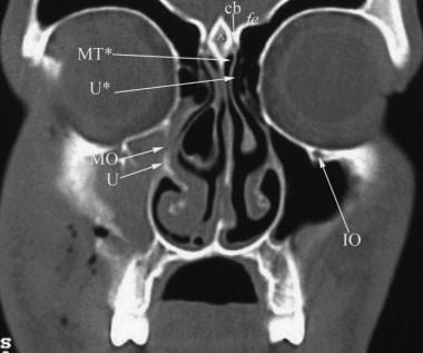


Ct Scan Of The Paranasal Sinuses History Basic Concepts Anatomy
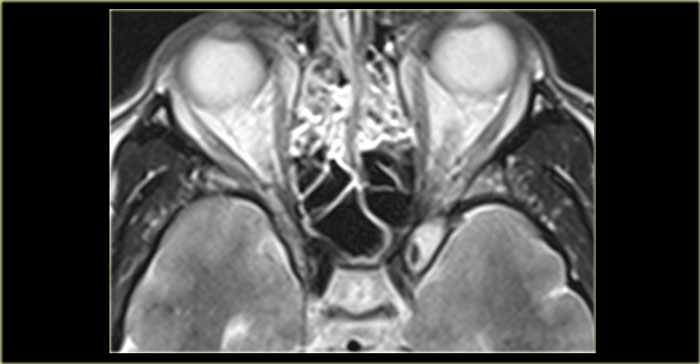


The Radiology Assistant Mri Examination
Of the paranasal sinuses, the clinical si gnificance of anatomical variants, and the terminology used in functional endoscopic sinus surgery is basic to the correct interpretation of imagingThis MRI orbits and paranasal sinuses cross sectional anatomy tool is absolutely free to use Use the mouse scroll wheel to move the images up and down alternatively use the tiny arrows (>>) on both side of the image to move the images>>) on both side of the image to move the imagesTemporal Bone Anatomy 10;
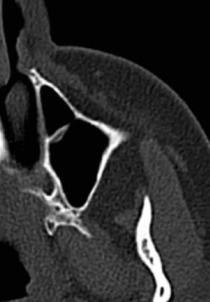


Paranasal Sinus Anatomy What The Surgeon Needs To Know Intechopen


The Preoperative Sinus Ct Avoiding A Close Call With Surgical Complications Radiology
Overall, the most common anatomic variant of the paranasal sinuses and nasal cavity was nasal septal deviation It was present to some extent in 1 of 192 patients (984%) but was considered to be more than minimal (> 1 mm) in 118 of 192 patients (614%) The second most common variant was Agger nasi cells, which were present in 160 of 192 patients (3%) The third most commonSinonasal anatomy Kubal WS(1) Author information (1)Department of Radiology, Medical College of Virginia, Virginia Commonwealth University, Richmond, Virginia , USA Recent advances in paranasal sinus imaging have been driven by the development of functional endoscopic sinus surgery The goal of most sinus imaging is to provide aDepartment of Radiology, University of Iowa Hospitals and Clinics, tomography are important supplements to patient history and physical examination in the diagnosis of diseases of the paranasal sinuses Background historical information regarding these radiologic modalities is presented, and the development and anatomy of the sinuses and roentgenographic views are


Paranasal Sinuses Annotated Ct Radiology Case Radiopaedia Org


Www Ijcmr Com Uploads 7 7 4 6 Ijcmr 1967 V4 Pdf
· Radiologic evaluation of the paranasal sinuses is essential in delineating the location and extent of sinonasal diseases and in planning surgical intervention Plain radiography, computed tomography and magnetic resonance imaging are applied in evaluating the sinusesThe paranasal sinuses usually consist of four paired airfilled spaces They have several functions of which reducing the weight of the head is the most important Other functions are air humidification and aiding in voice resonance They are named for the facial bones in which they are locatedRadiology of the Nasal Cavity and Paranasal Sinuses CT scan evaluation of the anatomical variations of the ostiomeatal complex 27 September 09 Indian Journal of Otolaryngology and Head & Neck Surgery, Vol 61, No 3 Appropriate interslice gap for screening coronal paranasal sinus tomography for mucosal thickening
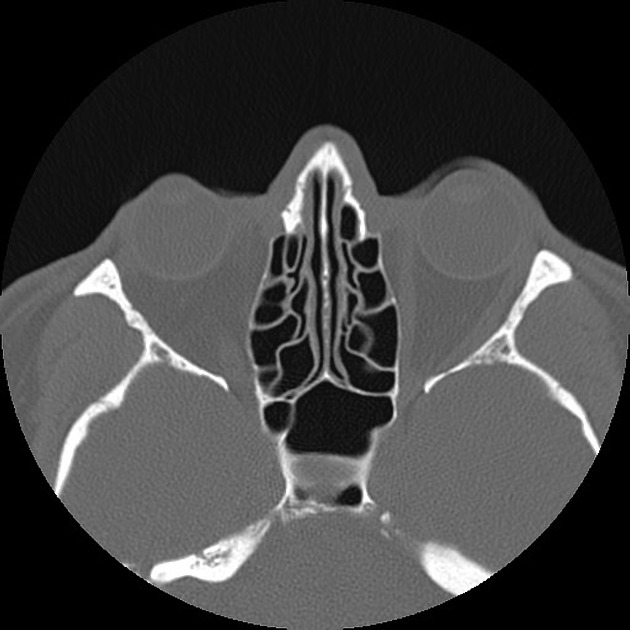


Normal Sinus Ct Annotated Radiology Case Radiopaedia Org
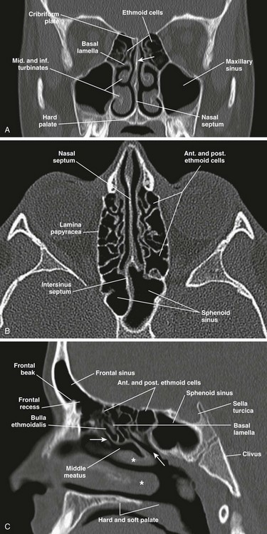


Nose And Sinonasal Cavities Radiology Key
· When it comes to imaging of neoplasms of the paranasal sinuses, CT and MRI play complementary roles It is not about the histology but about answering the question 'is it tumor or not?' and then determining the extent of the disease, for example intracranial or orbital extension Use MRI to differentiate inspissated secretions from neoplasmsSwallowing Swallowing disorders update;Department of Radiology, Mount Sinai Medical Center, New York, New York ABSTRACT As has happened in all facets of neuroimaging, crosssectional imaging has dramatically changed our approach and understanding of the anatomy and pathology of paranasal sinuses We have moved away from plain film radiographs to modern highresolution sinus computerized tomography


Q Tbn And9gcrsyvvumm9lju5sjjekqgfmg7lptxk 0fh0if 8fws5xzamqodp Usqp Cau


Paranasal Sinus Anatomy What The Surgeon Needs To Know Intechopen
Sinus CT is frequently requested by ear, nose and throat (ENT) specialists The CT test is usually made to evaluate the anatomy of the paranasal sinuses Information about the sinus anatomy of individual patients is essential prior to a FESS procedure (functional endoscopic sinus surgery)Frontal sinus, sphenoidal sinus,;Imaging Paranasal sinuses DRE 3 Dr Mamdouh Mahfouz Watch later Share Copy link Info Shopping Tap to unmute If playback doesn't begin shortly, try restarting your device


Atlas Of Imaging Of The Paranasal Sinuses Second Edition 2nd Editio


Orbits Anatomy Mri Orbits And Paranasal Sinuses Anatomy Free Cross Sectional Anatomy
Tinnitus Pulsatile and nonpulsatile tinnitus;The knowledge of the presence of the paranasal sinuses dates back to early mankind as well as attempts to treat their diseases Apart from the sensory function of smell, however, little has been known about the function and especially the anatomy of the system till the end of the last century Until the late middle ages sometimes obscure functions were attributed to the sinuses, like · Radiology Assistant app;


Computed Tomography Anatomy Of The Paranasal Sinuses And Anatomical Variants Of Clinical Relevants In Nigerian Adults Sciencedirect
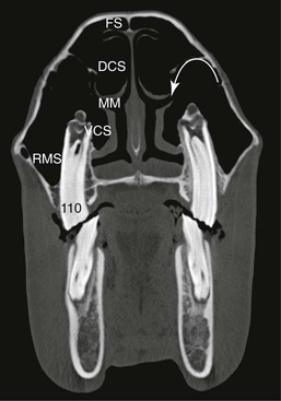


Diseases Of The Nasal Cavity And Paranasal Sinuses Veterian Key
· All radiologists should be familiar with the 3D anatomy of the paranasal sinuses and the anatomic variants that surgeons are likely to encounter This article reviews the embryology of the paranasal sinuses and outlines the CT technique/protocols for imaging this region CT anatomy of the nasal cavity and paranasal sinuses is described in detail together with theSurgical Anatomy of the Paranasal Sinuses Brent A Senior, MD, FACS, FARS Sheila and Nathaniel Harris Professor of Otolaryngology/Head and Neck Surgery and Neurosurgery University of North Carolina at Chapel Hill Disclosures • Consultant » Sinuwave » Laurimed » Entrigue » Nasoform » Olympus Gyrus • Speaker » Stryker » Genentech • Stockholder » Entrigue » Remedease »Analysis of every routine CT scan of the paranasal sinuses obtained for sinusitis or rhinitis for the presence of different anatomic variants is of questionable value unless surgery is planned Keywords anatomic variants, CT, functional endoscopic sinus surgery, paranasal sinuses, sinusitis
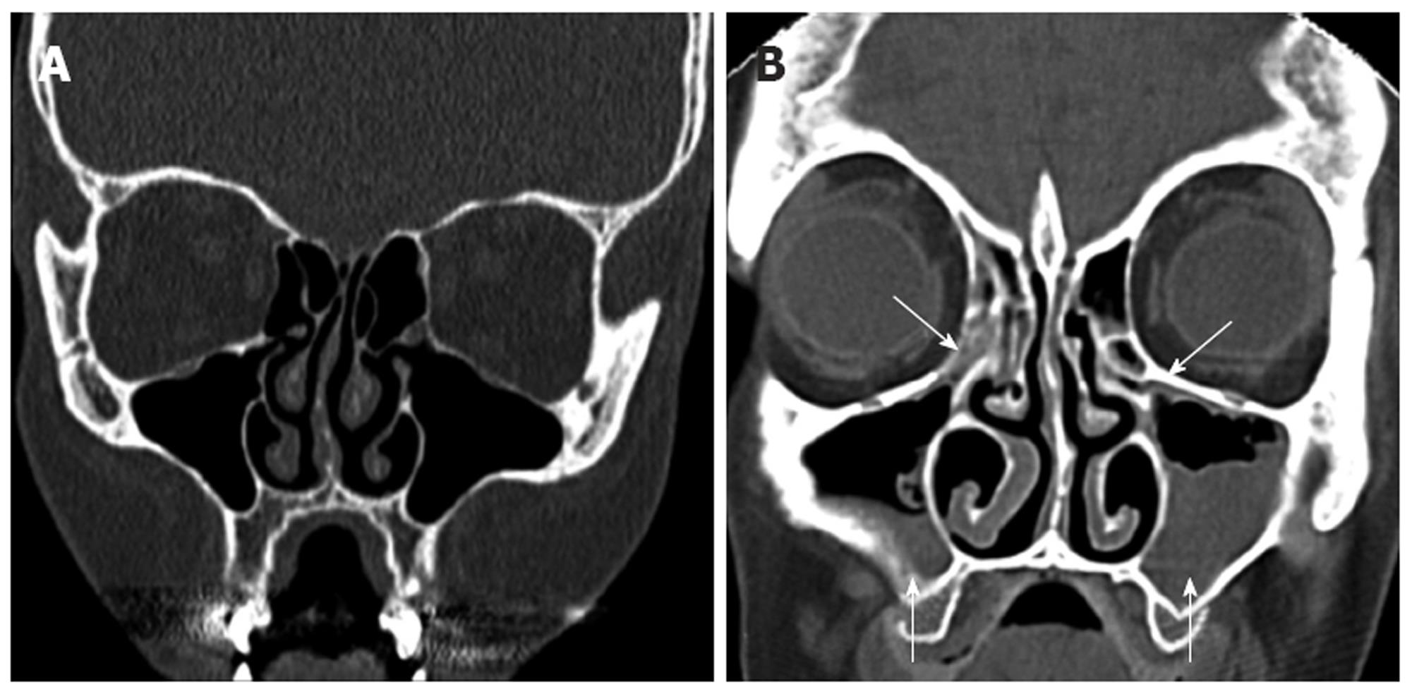


Computed Tomography Scans Of Paranasal Sinuses Before Functional Endoscopic Sinus Surgery
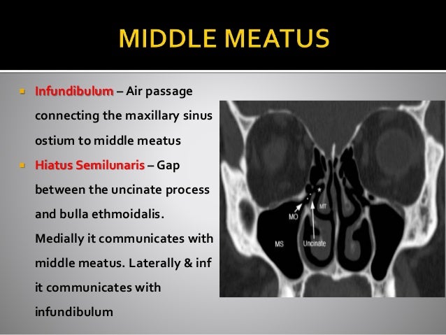


Ct Anatomy Of Para Nasal Sinuses


Exam 4 Anatomy And Diseases Of The Paranasal Sinuses Flashcards Quizlet


Startradiology


Imaging Of Paranasal Sinuses And Anterior Skull Base And Relevant Anatomic Variations Radiologic Clinics


The Frontal Sinus Drainage Pathway And Related Structures American Journal Of Neuroradiology


Ct And Mri Show Complexparanasal Sinus Anatomy


Paranasal Sinuses And Nasopharynx Ct And Mri European Journal Of Radiology


21 Best Paranasal Sinuses Ideas Paranasal Sinuses Sinusitis Radiology


Imaging Of The Paediatric Paranasal Sinuses Document Gale Onefile Health And Medicine


Computed Tomography Anatomy Of The Paranasal Sinuses And Anatomical Variants Of Clinical Relevants In Nigerian Adults Sciencedirect


A Ct Scan In Coronal Plane Shows The Normal Paranasal Sinus Anatomy Download Scientific Diagram


Normal Ct Paranasal Sinuses Radiology Case Radiopaedia Org


The Canine Head And Skull Ct Atlas Of Veterinary Clinical And Radiological Anatomy Of The Dog


Paranasal Sinus Disease Ento Key
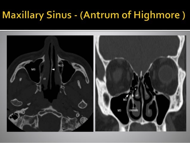


Ct Anatomy Of Para Nasal Sinuses


Orbits Anatomy Mri Orbits And Paranasal Sinuses Anatomy Free Cross Sectional Anatomy
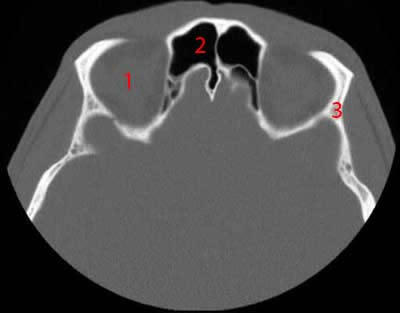


Paranasal Sinuses Ct Anatomy W Radiology


Of The Paranasal Sinuses Radiology Key
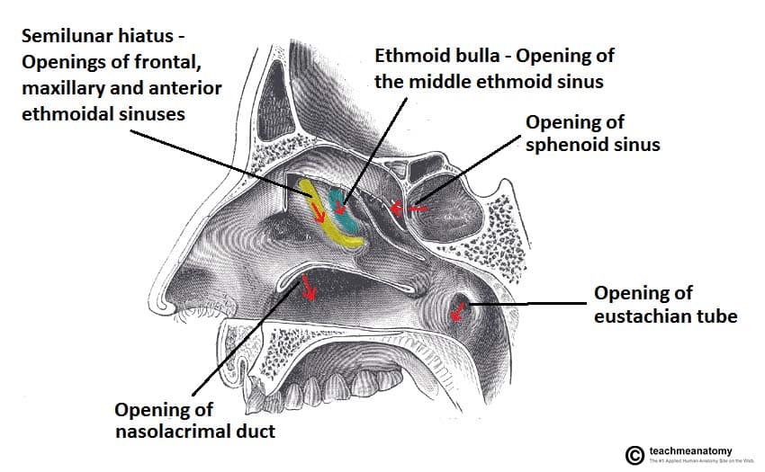


The Paranasal Sinuses Structure Function Teachmeanatomy


Q Tbn And9gcskgu9etd0groaaluz7emwelm1ldaxh Hayt6npc2co5xizzbre Usqp Cau


Radiology Of The Nasal Cavity And Paranasal Sinuses Ento Key


Pin On Paranasal Sinuses
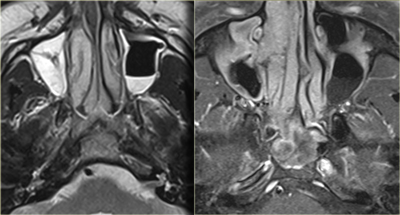


The Radiology Assistant Mri Examination


Normal Rabbit Paranasal Sinuses The Maxillary Sinus Ostium Is Seen Download Scientific Diagram
:background_color(FFFFFF):format(jpeg)/images/article/en/the-paranasal-sinuses/972PC0nYOzlz7wqSgLmNA_sinus_frontalis_large_u9Vfozc0uUoMtc6KtIaUfw.png)


Paranasal Sinuses Anatomy And Clinical Aspects Kenhub


Epos C 093


Normal Anatomy And Anatomic Variants Of The Paranasal Sinuses On Computed Tomography Neuroimaging Clinics


Www Birpublications Org Doi Pdf 10 1259 Dmfr 1905


Computed Tomography Anatomy Of The Paranasal Sinuses And Anatomical Variants Of Clinical Relevants In Nigerian Adults Sciencedirect


Pin On Anatomia
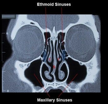


Sinus Anatomy Sinushealth Com


William T O Brien Sr Incredibly Grateful To Be A Part Of This Program Esp Covering One Of My Favorite Topics Paranasal Sinuses Promise For It To Be Image Rich
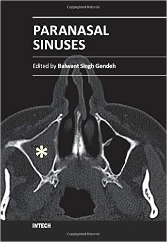


Paranasal Sinuses Medicine Health Science Books Amazon Com


Basic Ct Anatomy Of Paranasal Sinuses Made Easy Youtube


Ct Scan Of Paranasal Sinus Axial Coronal Youtube
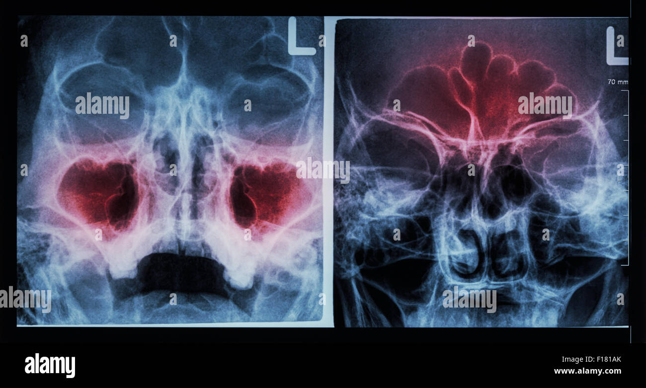


Film X Ray Paranasal Sinus Show Sinusitis At Maxillary Sinus Left Image Frontal Sinus Right Image Stock Photo Alamy


The Radiology Assistant Paranasal Sinuses Mri


Http Pdf Posterng Netkey At Download Index Php Module Get Pdf By Id Poster Id


Orbits Anatomy Mri Orbits And Paranasal Sinuses Anatomy Free Cross Sectional Anatomy


1


Agenesis Of Paranasal Sinuses And Nasal Nitric Oxide In Primary Ciliary Dyskinesia European Respiratory Society


Pin On Anatomia


Paranasal Sinus Imaging European Journal Of Radiology
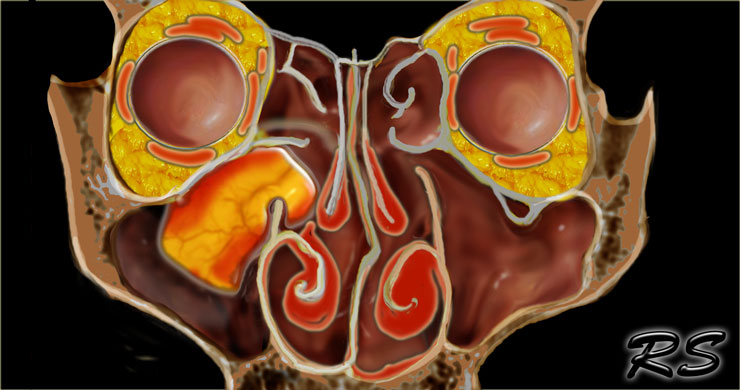


The Radiology Assistant Mri Examination


Startradiology


Ct Of Paranasal Sinuses Stock Image P410 0085 Science Photo Library


Radiologic Imaging In The Management Of Sinusitis American Family Physician
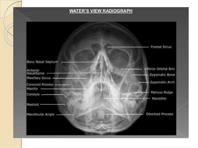


Radiology Of Nose And Paranasal Sinuses
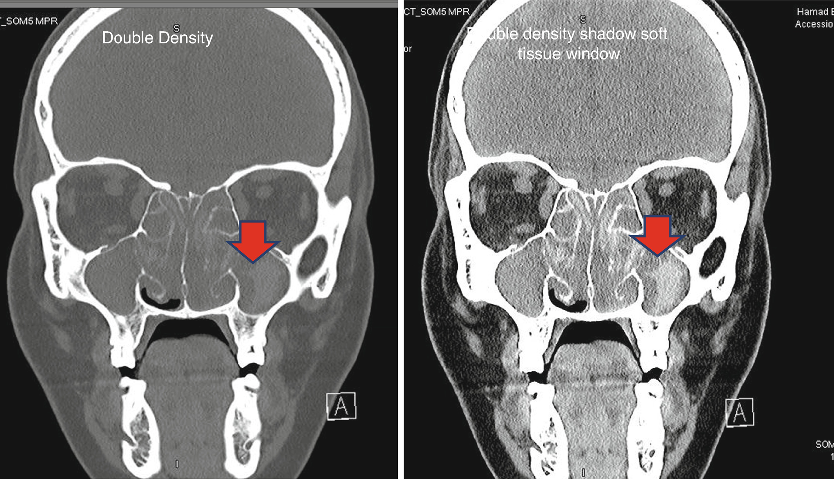


Radiology Of Paranasal Sinuses Springerlink


Home Page


Overview Of The Drainage Pathways Of The Paranasal Sinuses A Download Scientific Diagram


Paranasal Sinus Anatomy What The Surgeon Needs To Know Intechopen


Figure 5 From Paranasal Sinus Anatomy What The Surgeon Needs To Know Semantic Scholar


Http Pdf Posterng Netkey At Download Index Php Module Get Pdf By Id Poster Id


A E Coronal Axial And Sagittal Ct Images Of The Bone Window Demonstrate The Normal Anatomy Of The Paranasal Sinuses And The Integrity Of The Nasal Septum Nasal Bone And Nasal Pyramid


The Preoperative Sinus Ct Avoiding A Close Call With Surgical Complications Radiology


The Preoperative Sinus Ct Avoiding A Close Call With Surgical Complications Radiology


Www Ajronline Org Doi Pdf 10 2214 Ajr 14


Cureus Paranasal Sinus Wall Erosion And Expansion In Allergic Fungal Rhinosinusitis An Image Scoring System


Startradiology


Q Tbn And9gcthldxvr6nykftmuawhyfamnfge8pfsjllwgh3teyo Usqp Cau


Researchers Cast New Light On Equine Sinuses Horsetalk Co Nz


The Frontal Sinus Drainage Pathway And Related Structures American Journal Of Neuroradiology


Ct Sinus Anatomy Anatomy Drawing Diagram


Cone Beam Computed Tomography For The Nasal Cavity And Paranasal Sinuses Dental Clinics
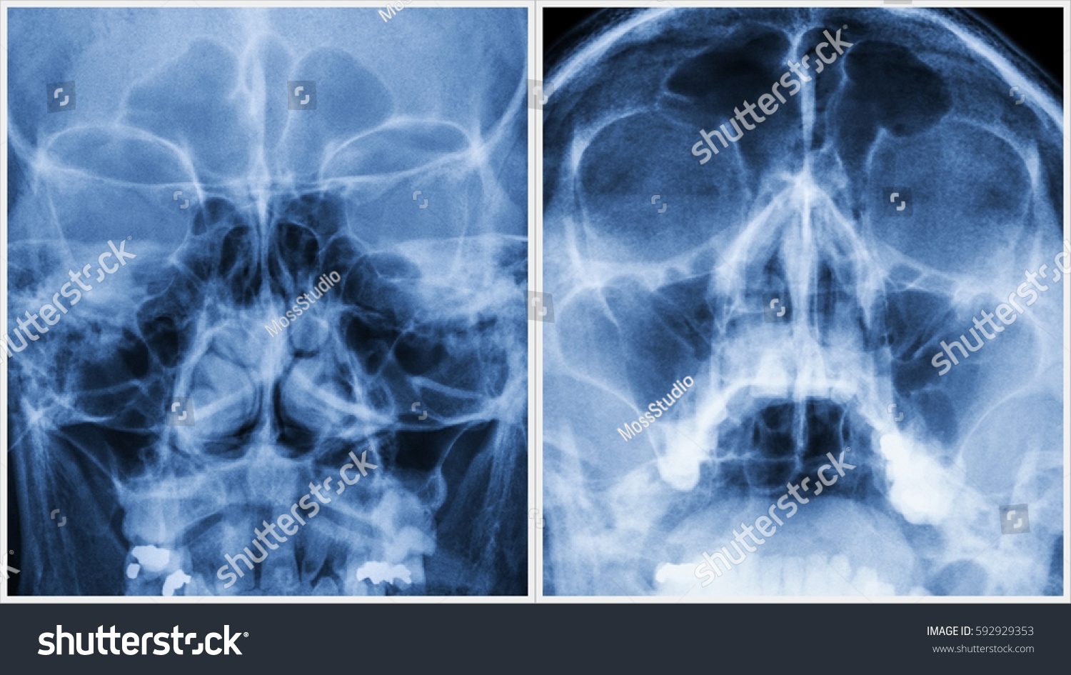


Film Xray Paranasal Sinuses Pns Caldwell Stock Photo Edit Now


Pictorial Essay Anatomical Variations Of Paranasal Sinuses On Multidetector Computed Tomography How Does It Help Fess Surgeons Abstract Europe Pmc
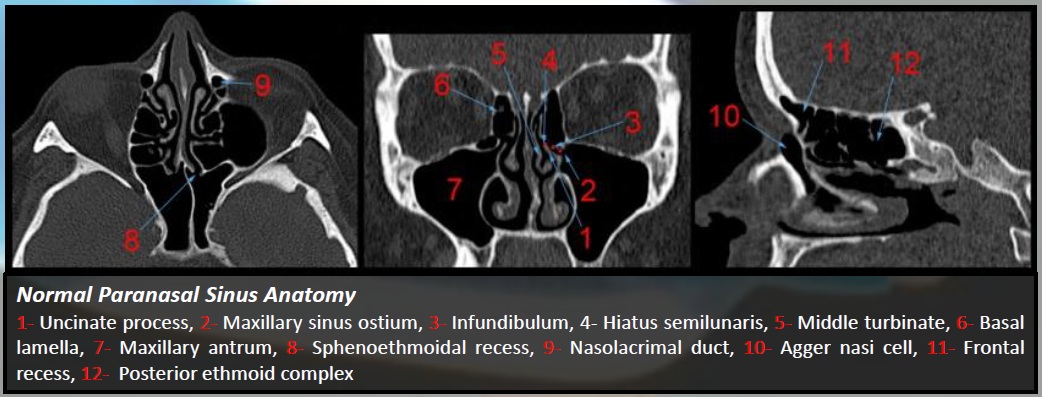


Epos C 0793
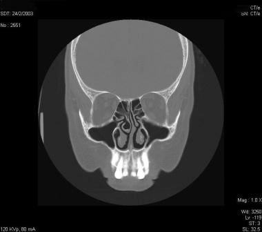


Sinusitis Rhinosinusitis Imaging Practice Essentials Radiography Computed Tomography


Imaging Of The Paranasal Sinuses And Nasal Cavity Normal Anatomy And Clinically Relevant Anatomical Variants Sciencedirect
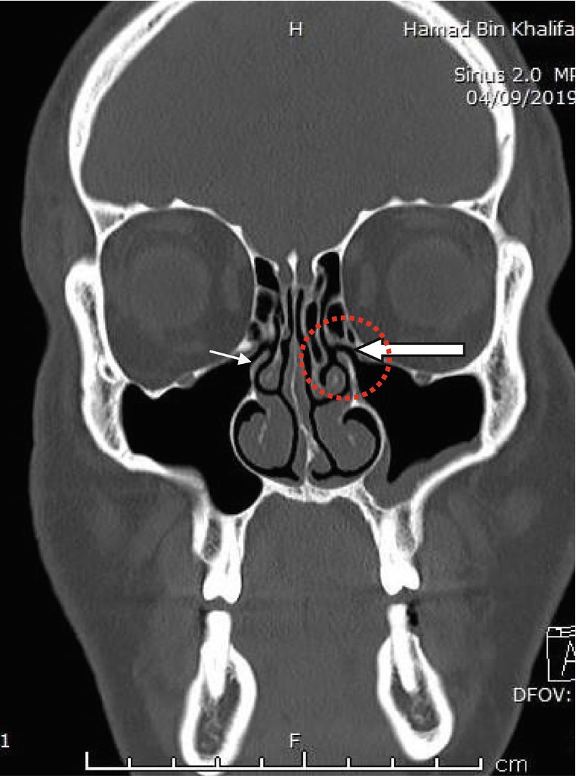


Radiology Of Paranasal Sinuses Springerlink


Sinusitis Cancer Therapy Advisor


Paranasal Sinuses Radiography W Radiology


コメント
コメントを投稿